You already sat through the slide with dendrites, soma, axon, and terminal buttons. This reading isn't going to redraw that diagram for you. What it will do is connect the dots your slides leave implicit: why the wiring is built the way it is, where students usually get confused, and what it means when a headline says a brain scan "found" the seat of love, addiction, or free will.
One neuron, one decision
Picture the neuron the way your slide laid it out: dendrites gather input, the soma (cell body) integrates it, the axon carries the outgoing signal, and the terminal buttons pass it along. What the slide's static image can't show you is that this is a live decision process happening constantly, all over your nervous system, in parallel, thousands of times a second.
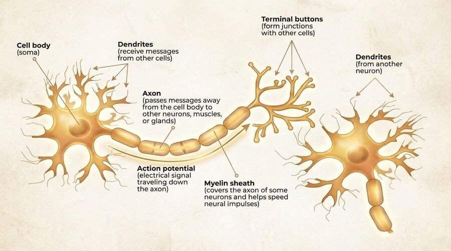
Here's the part worth sitting with. The action potential — the electrical signal that travels down the axon — is all-or-none. A neuron doesn't fire "a little bit" in response to a weak stimulus and "a lot" in response to a strong one. Either the input reaching the soma crosses threshold and the neuron fires a full-strength spike, or it doesn't fire at all. There's no dial, only a switch. What varies with stimulus intensity isn't the size of any single spike — it's the rate of firing and how many neurons join in. A bright light doesn't give you a bigger action potential than a dim one; it gives you more of them, faster, across more cells.
It's worth knowing what the spike actually is at the level of ions, because the mechanism explains the switch-like behavior. At rest, a neuron holds a small negative voltage across its membrane — around −70 millivolts — maintained by an uneven distribution of charged particles, with more sodium ions outside the cell and more potassium inside. This is the resting potential: a charged, ready state, like a cocked mousetrap. When incoming signals push the voltage up past a critical threshold (roughly −55 mV), voltage-gated channels snap open and sodium rushes in, flipping the local voltage sharply positive — that's depolarization, the action potential itself. Channels then reverse to pump the neuron back to rest (repolarization), and for a brief refractory period the cell cannot fire again no matter how strong the input. That refractory window is part of why signals travel in one direction and why there's a ceiling on how fast a neuron can fire (Bear et al., 2020).
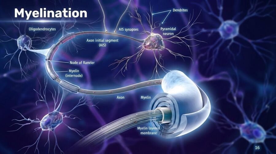
Myelin — the fatty sheath wrapped around many axons — is what makes this fast enough to be useful. It insulates the axon and lets the signal jump between the gaps (nodes of Ranvier) instead of crawling down every inch of membrane. This is also not fixed at birth: myelination is experience-dependent, meaning the sheathing around circuits you actually use gets reinforced over time. Skill practice, in a very literal sense, is partly an insulation project.
Three kinds of neuron, and the reflex that skips the brain
The diagram makes every neuron look the same, but neurons come in three functional flavors that your slides name and your exam will expect. Sensory neurons carry information toward the central nervous system — from your skin, eyes, ears, and organs inward. Anatomists call these afferent fibers; a handy mnemonic is that afferent signals arrive. Motor neurons run the other way, carrying commands from the CNS out to muscles and glands — efferent fibers, which exit. And interneurons, which are the overwhelming majority of neurons you own, live entirely inside the brain and spinal cord and connect everything to everything else. Every nerve in your body is a bundle of thousands of axons running both directions at once, a two-way superhighway.
The clearest demonstration that these three types are genuinely different is the spinal reflex. Touch a hot stove and your hand is already moving before you feel the burn. That's because the sensory neuron fires into the spinal cord, an interneuron there hands the signal straight to a motor neuron, and the motor neuron yanks the arm — all without waiting for the message to climb to the brain and back. The brain finds out a fraction of a second later, which is why the "ouch" arrives after the flinch. The knee-jerk your doctor tests is the same architecture with even fewer steps. The spinal cord, in other words, isn't just the cable connecting brain to body; it's a smart relay with a few automatic programs of its own, and every segment of it can run one.
Myelin, from the previous section, is what makes all of this fast — and its loss shows how much rides on that insulation. In multiple sclerosis, an autoimmune disease, the immune system strips myelin from axons throughout the nervous system. The signal doesn't stop, but it slows and stutters, and the result is the fatigue, loss of motor control, dizziness, and other symptoms of the disease. Nothing is wrong with the neurons' ability to fire; the problem is entirely in the wiring's insulation.
Electrical within, chemical between
This is the distinction students blur most on exams, so it's worth making explicit and keeping separate in your head.
Within a neuron
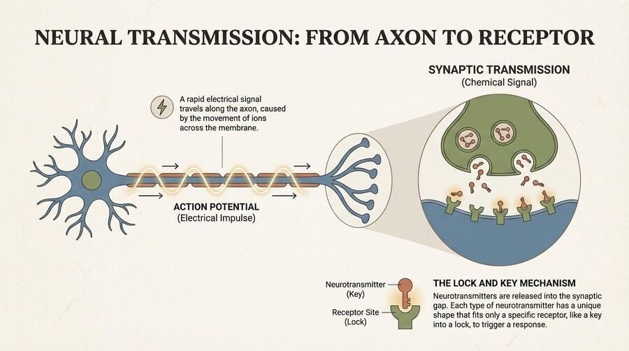
Signaling is electrical. The action potential is a wave of voltage change that travels down the axon, from soma to terminal buttons. One neuron, one continuous electrical event.
Between neurons
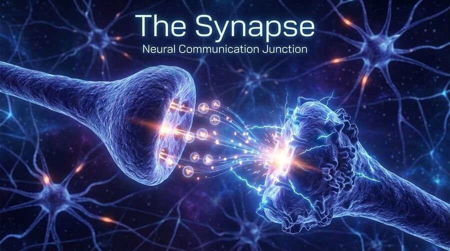
Signaling is chemical. When the electrical signal reaches the terminal buttons, it triggers the release of neurotransmitters into the synapse — the tiny gap between neurons. Those chemicals cross the gap, bind to receptors on the next neuron, and either excite or inhibit it. Afterward, leftover neurotransmitter is cleared out largely through reuptake, where the sending neuron reabsorbs it for reuse.
Why it matters: this chemical step — release, binding, reuptake — is exactly where psychoactive drugs do their work. An agonist mimics or boosts a neurotransmitter's effect at the receptor; an antagonist blocks it. A drug that slows reuptake leaves more neurotransmitter sitting in the synapse longer, amplifying its effect. None of this touches the electrical action potential directly — it all happens at the chemical handoff between cells. That's also why so much of pharmacology, and so much of clinical psychology's biological toolkit, is really synapse-level engineering.
One more piece makes sense of why the neuron behaves like a decision-maker rather than a relay. A single neuron receives input from thousands of others at once, and those inputs are not all pushing the same way. Some neurotransmitters produce excitatory effects that nudge the receiving cell toward firing; others produce inhibitory effects that push it away from firing. The cell continuously adds these up — a process called summation — and only fires if the excitatory input outweighs the inhibitory input by enough to cross threshold. So every spike is really the outcome of a tiny, constantly re-run vote among incoming signals. That is what people mean when they say the neuron "integrates" information rather than merely passing it along.
The major neurotransmitters, briefly
Your slide likely listed these. Here's what each one is mainly doing, in a sentence apiece, so the names stop being interchangeable jargon:
Dopamine — reward, motivation, and movement. Serotonin — mood, sleep, and appetite regulation. GABA — the brain's main inhibitory neurotransmitter, calming activity down. Glutamate — the main excitatory neurotransmitter, the accelerator to GABA's brake. Acetylcholine — muscle movement and memory formation. Norepinephrine — arousal and alertness. Endorphins — the body's own pain relief and pleasure signaling.
These names stop feeling like trivia once you see them attached to conditions and drugs you already know. The loss of dopamine-producing cells in one brain pathway is central to Parkinson's disease, which is why its motor symptoms are treated with drugs that boost dopamine. Many antidepressants (the SSRIs) work by blocking serotonin reuptake, leaving more of it in the synapse. Anti-anxiety drugs like benzodiazepines enhance GABA, dialing down neural excitability — the same calming system alcohol acts on. Acetylcholine is the transmitter that tells muscles to contract, which is exactly why toxins that block it, such as botulinum (Botox) or curare, cause paralysis; its decline is also implicated in the memory loss of Alzheimer's disease. And endorphins occupy the same receptors that opioid drugs like morphine and heroin hijack — those drugs feel powerful precisely because they impersonate a signaling system the body already uses for pain and reward.
That opioid story has a life-saving footnote your slides highlight. Naloxone (Narcan) is a pure antagonist at the opioid receptor: it fits the receptor better than heroin or fentanyl does, shoves them off, and activates nothing — which is why a single nasal spray can reverse an overdose in minutes, restoring breathing that the drug had shut down. Methadone works on the opposite logic: it's a slow, long-acting agonist that keeps the receptors steadily occupied, so a person in treatment avoids withdrawal without the rapid spike that produces a high. Both are the agonist/antagonist distinction turned into medicine. Toxicologists also separate acute toxicity — the harm one dose can do right now, like an overdose stopping breathing — from chronic toxicity, the damage that accumulates from repeated use over months or years. A drug can be low on one and high on the other.
◆ Update: the "chemical imbalance" story is oversimplified
You've probably heard depression explained as "a chemical imbalance — too little serotonin." That framing is a slogan, not a settled finding. A large umbrella review of the evidence found that the data do not support the simple claim that low serotonin causes depression (Moncrieff et al., 2023). Neurotransmitters clearly matter to mood — that's not in dispute — but the tidy, single-cause story oversells a mechanism science hasn't actually nailed down. This is a good example of how a metaphor that's easy to put on a pamphlet can outrun the evidence behind it.
Zooming out: how the brain is organized
Your slides walked bottom-to-top through brain structure, and that ordering isn't arbitrary — it roughly tracks evolutionary age and how automatic versus deliberate the functions are.
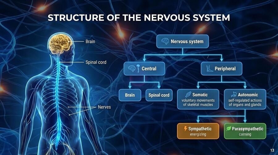
Before zooming in on the brain itself, it helps to place it in the larger system the diagram lays out. The central nervous system (CNS) is the brain and spinal cord — the command center and its main cable. Everything else is the peripheral nervous system (PNS), the network of nerves carrying messages to and from the rest of the body. The PNS in turn divides in two. The somatic branch handles voluntary movement and carries sensory information inward. The autonomic branch runs the involuntary machinery — heart rate, digestion, pupil size — and it has two opposing halves that are worth memorizing as a pair. The sympathetic nervous system is the accelerator: it mobilizes the body for "fight or flight," flooding you with energy, speeding the heart, and dilating the pupils when you're startled or stressed. The parasympathetic nervous system is the brake: it handles "rest and digest," slowing you back down and restoring the body once the threat has passed. Most of the time these two are in a moving balance, which is why you can go from calm to alarmed and back within a single minute.
The nervous system isn't the body's only messaging network, either. Working alongside it, and more slowly, is the endocrine system — a set of glands that release hormones directly into the bloodstream. Where neural signals are fast and precise (milliseconds, targeted to specific cells), hormonal signals are slower and more diffuse, traveling everywhere the blood goes and producing effects that can last minutes, hours, or longer. The two systems are deeply intertwined: the hypothalamus, at the base of the brain, steers the pituitary gland, which in turn directs other glands. When you're frightened, for instance, the adrenal glands dump adrenaline and cortisol into the blood, sustaining the very "fight or flight" state the sympathetic nervous system kicked off electrically. Fast system to start it, slow system to keep it going.
At the base, the brainstem and hindbrain handle the functions you don't have to think about and can't easily override: heartbeat, breathing, arousal, basic survival. Above that sits the limbic system, where the amygdala handles threat detection and emotional reactivity and the hippocampus handles forming new memories. On top of all of it is the cerebral cortex, wrapped in its four lobes: the frontal lobe for control, planning, and decision-making; the parietal lobe for touch and spatial processing; the temporal lobe for hearing and language; and the occipital lobe for vision.
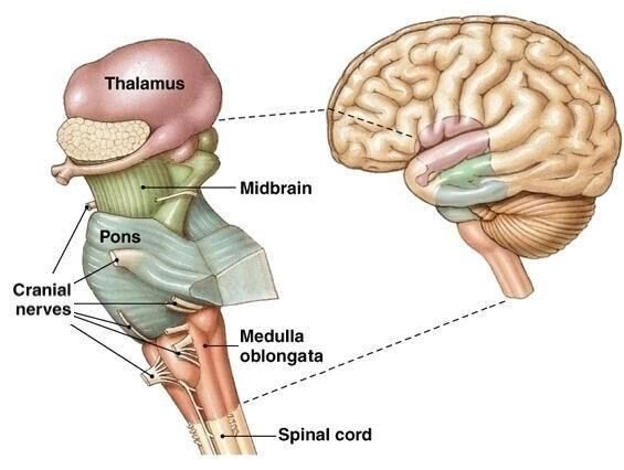
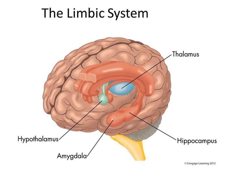
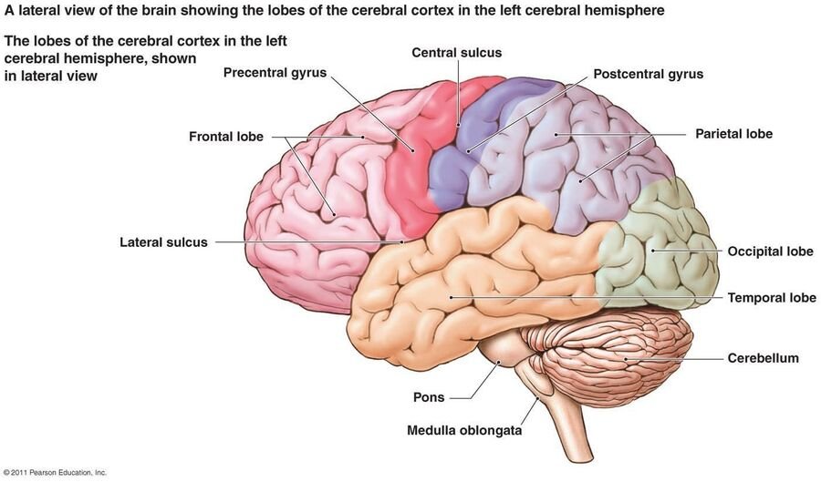
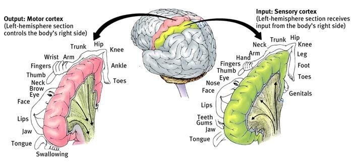
One structure the diagrams show but the lobe list skips is the cerebellum, the dense wrinkled ball tucked at the back beneath the cortex. Long thought to handle only balance, coordination, and the timing of smooth movement, it's now known to contribute to some cognitive and language functions as well — a reminder that the brain's division of labor is rarely as clean as a labeled diagram suggests.
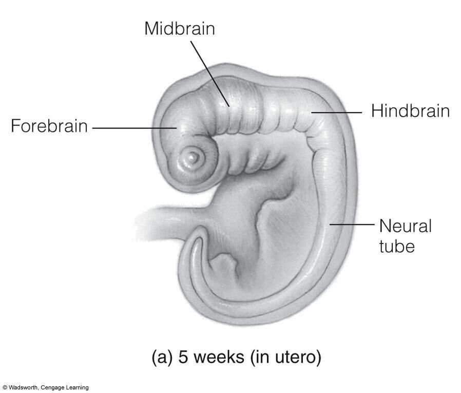
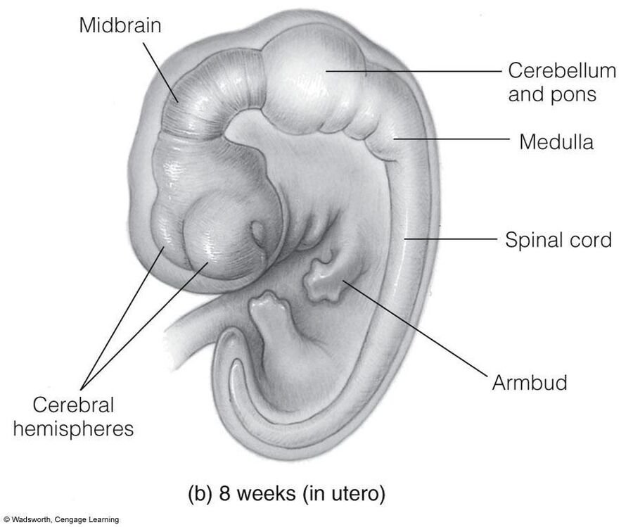
Two ways of carving up the same organ are worth holding side by side, because exams use both. The developmental scheme, visible in the embryo figures below, divides the brain into hindbrain, midbrain, and forebrain. The hindbrain is the evolutionarily oldest part and runs survival: the medulla oblongata keeps your heart beating and your lungs breathing without your permission; the pons (literally "bridge") connects the hindbrain to the rest of the brain and helps regulate sleep, especially REM; the cerebellum sits beside them. The midbrain is a small relay for sensory and motor traffic, and threading up through both is the reticular formation (or reticular activating system), a net of neurons that controls arousal — it's the dial between coma, sleep, and alert wakefulness, and damage to it can leave a person permanently unconscious. The forebrain is the newest and largest division, and in humans it has ballooned: it holds the cortex plus the subcortical structures underneath it. Three of those deserve names. The thalamus is the sensory switchboard — every sense except smell is routed through it on the way to the cortex. The hypothalamus, tiny and just below it, maintains homeostasis (temperature, hunger, thirst, blood pressure) and drives the basic motivations lecture summarizes as the "Four F's": fighting, fleeing, feeding, and mating. And the amygdala and hippocampus, from the limbic list above, round out the set. This is an expensive machine: your brain weighs about three pounds, roughly 2% of your body weight, and burns about 20% of your resting energy. Thinking is not free.
One more piece of cortical geography that the homunculus figure shows but doesn't spell out: the two "strips" face each other across the central sulcus, the groove that separates the frontal lobe from the parietal lobe. The motor cortex runs along the back edge of the frontal lobe and sends commands out to the muscles; the somatosensory cortex runs along the front edge of the parietal lobe and receives touch, temperature, and pain from the body. Each is a map of the body, laid out point for point, and each map is distorted the same way — the more precise the control or the finer the sensitivity, the more cortex the body part gets. Lips and fingertips are enormous; your back is a sliver. And because of the crossover you'll meet in the next unit, the left strip handles the right side of the body and vice versa.
This layered structure isn't fixed hardware, either. The brain shows neuroplasticity — it rewires itself with use, strengthening the connections you exercise and pruning the ones you don't. A striking illustration comes from London taxi drivers, who must memorize the city's tangle of streets to earn their license: Maguire et al. (2000) found that the rear of their hippocampus, a region central to spatial memory, was measurably enlarged compared with non-drivers, and grew larger the longer they had been driving. The structure had physically adapted to the demand placed on it. Myelination getting reinforced with practice, mentioned earlier, is another expression of the same broader principle: the brain you have today is shaped, down to its anatomy, by what you've actually been doing with it.
The myth
We only use 10% of our brain — the rest is untapped potential just waiting to be unlocked.
What's actually true
Virtually all of the brain is active over the course of a day. Brain imaging doesn't show 90% of the brain sitting dark. What's true is that not every region is maximally active at the exact same moment — different tasks recruit different networks. "Not all at once" got flattened into "only 10% at all," and the flattened version stuck around because it's a great movie premise.
Watching the brain work: methods and their tradeoffs
Your slides likely named several imaging and manipulation techniques. Each one buys you something and costs you something else — mainly a tradeoff between when something happened and where.
Step 1 · EEG
Electrodes on the scalp track electrical activity in real time. Excellent temporal resolution — it can catch changes millisecond to millisecond — but poor spatial resolution. It tells you something happened right now; it's much vaguer about exactly where.
Step 2 · CT and MRI
These image brain structure — the physical anatomy — rather than ongoing activity. Useful for spotting damage, tumors, or structural differences, but they're a snapshot of the hardware, not a recording of the brain in action.
Step 3 · PET and fMRI
These aim to capture brain activity. fMRI in particular offers good spatial resolution — it can localize activity fairly precisely — but poor temporal resolution, and, critically, it's indirect. fMRI doesn't measure neurons firing. It measures blood oxygenation — the BOLD signal — as a proxy for neural activity, on the assumption that busier neurons draw more oxygenated blood. That's a reasonable assumption, but it's an inference, not a direct readout of the brain thinking (Logothetis, 2008).
Step 4 · TMS
Transcranial magnetic stimulation nudges neural activity in a targeted region up or down. Because it manipulates rather than just observes, it's one of the few tools that lets researchers move toward causal claims — if disrupting a region disrupts a function, that's stronger evidence than just watching the region light up during that function.
A few details the stepper compresses. CT is a stack of X-rays reassembled by computer into a cross-section — cheap and fast, good for spotting a tumor or bleeding, and it uses radiation. PET also uses radiation, but differently: the person is injected with a mildly radioactive tracer that rides the bloodstream, and because busier regions draw more blood, the tracer pools where the brain is working. That gives you activity rather than anatomy, but blurry and slow, and at the cost of a dose of radiation — which is why fMRI has largely replaced PET for research, except where PET's ability to tag specific neurotransmitter receptors is the whole point. MRI uses a strong magnetic field and no radiation at all, and fMRI is the same machine watching blood oxygenation change over time.
Your lecture gives you four dimensions for judging any of these tools, and it's a useful checklist to run on any brain study you read. Spatial resolution: how precisely can it say where? Temporal resolution: how precisely can it say when? Invasiveness: does it inject, irradiate, or open anything, or is it a cap on the scalp? Cost: an EEG rig is affordable for a small lab; an fMRI scanner is a multimillion-dollar magnet in a shielded room. No single method wins on all four. EEG wins on time, invasiveness, and cost but loses on space; fMRI wins on space and loses on time and cost; PET is invasive and expensive but sees chemistry the others can't. Choosing a method is choosing which of these you're willing to give up.
◆ The caveat that matters most
Most neuroimaging is correlational, not causal. A region "lighting up" on a scan during a task tells you that region was more active during that task — it does not make that region "the center for" love, honesty, or anything else, however catchy that headline sounds. Poldrack (2018) makes the case at length: neuroimaging can tell us a great deal, but reverse-inferring a specific mental state from a pattern of activation is far shakier than popular coverage implies.
The point gets made vividly — and a little absurdly — by the famous dead-salmon study. Researchers ran a dead Atlantic salmon through an fMRI task and, without correcting for the huge number of statistical comparisons involved in whole-brain imaging, found "significant" activation. The salmon was dead. The result was noise, not neural life (Bennett et al., 2010). It's a demonstration of why multiple-comparisons correction isn't optional bookkeeping — skip it, and you can find a "significant" brain response in a fish that isn't thinking about anything.
Bodhi says
When you read "a study found brain region X controls Y," pause and ask two questions: was this correlational (a scan during a task) or causal (a lesion, TMS, or intervention study)? And was it corrected for multiple comparisons? Those two questions alone will make you a more careful consumer of neuroscience headlines than most people writing them.
Check yourself
The one thing to carry out of this unit
Signaling within a neuron is electrical and all-or-none; signaling between neurons is chemical, and that chemical handoff is where drugs, disorders, and treatments mostly do their work. Zoom out and the brain's organization — brainstem, limbic system, cortex — tracks a rough gradient from automatic to deliberate. But however precise a scan looks, remember what it's actually measuring: mostly an indirect, correlational snapshot of activity, not a direct window into meaning. A lit-up region is a clue, not a verdict.
References
Bear, M. F., Connors, B. W., & Paradiso, M. A. (2020). Neuroscience: Exploring the brain (4th ed.). Jones & Bartlett.
Bennett, C. M., Baird, A. A., Miller, M. B., & Wolford, G. L. (2010). Neural correlates of interspecies perspective taking in the post-mortem Atlantic salmon: An argument for proper multiple comparisons correction. Journal of Serendipitous and Unexpected Results, 1(1), 1–5.
Logothetis, N. K. (2008). What we can do and what we cannot do with fMRI. Nature, 453(7197), 869–878.
Maguire, E. A., Gadian, D. G., Johnsrude, I. S., Good, C. D., Ashburner, J., Frackowiak, R. S. J., & Frith, C. D. (2000). Navigation-related structural change in the hippocampi of taxi drivers. Proceedings of the National Academy of Sciences, 97(8), 4398–4403. https://doi.org/10.1073/pnas.070039597
Moncrieff, J., Cooper, R. E., Stockmann, T., Amendola, S., Hengartner, M. P., & Horowitz, M. A. (2023). The serotonin theory of depression: A systematic umbrella review of the evidence. Molecular Psychiatry, 28(8), 3243–3256.
Poldrack, R. A. (2018). The new mind readers: What neuroimaging can and cannot reveal about our thoughts. Princeton University Press.