The lecture walks you from phrenology's bumps to Gazzaniga's flashing images. This page slows down on the two things students most often get backwards or oversimplify — how the visual wiring actually works, and what split-brain patients really tell us about consciousness — and then hands you the update your textbook probably hasn't caught up to yet.
From pseudoscience to a real idea
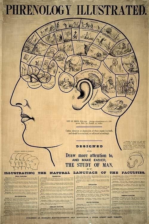
Phrenology, introduced by Franz Joseph Gall in the 1790s, was wrong about almost everything — you cannot read someone's character from skull bumps. But buried in the nonsense was a prescient idea that turned out to be right: that different mental functions live in different, specific parts of the brain. That's localization of function, and it's the thread connecting this whole unit. Phrenology had the right hypothesis and a hilariously wrong method — a useful reminder that a bold idea and good evidence are two different things.
Bodhi says
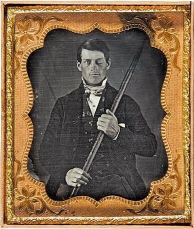
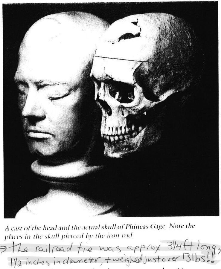
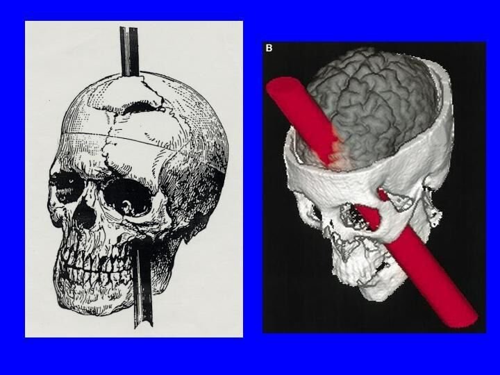
Phineas Gage is the case study version of the same point. A tamping iron through the frontal lobe didn't kill him — it changed his personality. That's the evidence that "who you are" isn't floating in some soul; it's tissue, and specific tissue at that. Gage is famous precisely because he's a natural experiment nobody would ever run on purpose.
◆ A caution about the Gage story
Gage is psychology's most retold case, and in the retelling he has often hardened into a cartoon — a hard-working gentleman transformed overnight into a violent, dissolute wreck. The historical record is more modest and more interesting than that. Macmillan's (2000) careful review of the original sources found that many of the lurid "facts" about Gage's later life were invented or embellished by writers who never met him, and that Gage likely recovered a good deal of function — later holding down a demanding job driving a stagecoach in Chile. The core lesson survives: damage to the frontal lobes can genuinely alter personality and self-control. But Gage is also a case study in how a striking story gets exaggerated each time it's retold, which is worth remembering every time a single dramatic case is offered as proof of anything.
The first hard evidence: Broca and Wernicke
Gage hinted that the frontal lobes mattered for personality, but the cleanest early proof that a specific mental function lives in a specific place came from the study of language. In 1861 the French physician Paul Broca examined a patient nicknamed "Tan" — the one syllable the man could still utter — who understood speech but had lost nearly all ability to produce it. When the patient died, Broca found damage to a particular region of the left frontal lobe, now called Broca's area. About a decade later, Carl Wernicke described the mirror-image deficit: patients with damage to a region of the left temporal lobe (now Wernicke's area) could speak fluently but produced jumbled, meaningless language and struggled to understand what was said to them. Put side by side, the two cases drew a startlingly precise map — one region for producing speech, a nearby one for comprehending it, both on the left. Modern high-resolution imaging of Broca's own preserved specimens has since confirmed and refined the picture, showing the damage in his patients extended somewhat beyond the small patch that bears his name (Dronkers et al., 2007). This was localization's founding evidence, and it is why, in the wiring section below, "language lives on the left" is not an arbitrary rule but a hard-won discovery.
Language is the famous case, but the right hemisphere has its own signature deficit, and it's stranger. Damage to the right parietal lobe — often from a stroke — can produce hemispatial neglect: the patient stops attending to the left half of the world. Not blindness; the eyes work. The person shaves or applies makeup to only the right side of the face, eats from only the right half of the plate and complains of being hungry, and, asked to copy a clock, crams all twelve numbers into the right half of the circle. Turn the plate around and they'll happily eat the rest. Neglect is localization's mirror image: whereas Broca's patients lose a function, neglect patients lose a region of space, which tells you the right parietal cortex is doing something like maintaining a map of where things are around you — and that the left hemisphere can't fully cover for it. It also gives you a vocabulary pair for the exam: each hemisphere mostly handles the contralateral (opposite) side of the body and space, not the ipsilateral (same) side, which is exactly why right-parietal damage erases the left.
The wiring rule everyone flips: fields, not eyes
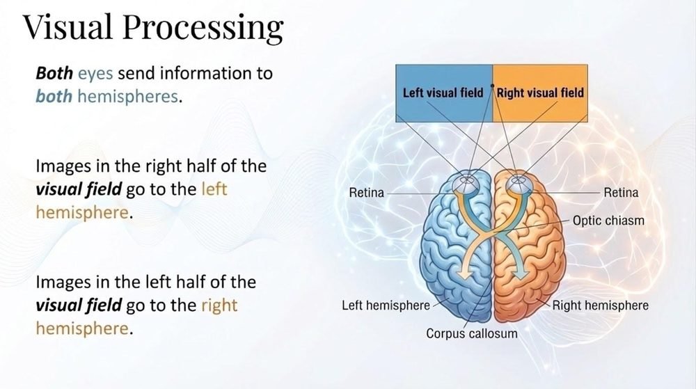
Here is the single most common mistake on this material. It is not "the left eye goes to the right brain." Each eye sends signals to both hemispheres. What crosses over is the visual field — the left and right halves of what you're looking at. Walk it through:
Step 1 · Two visual fields, not two eyes
Fix your gaze on a point. Everything to the left of that point is your left visual field; everything to the right is your right visual field. Both eyes see both fields.
Step 2 · The crossover
Because of how the optic nerves route at the optic chiasm, the right visual field → left hemisphere, and the left visual field → right hemisphere. Contralateral, like most of the body.
Step 3 · Language lives on the left
For most people, speech is a left-hemisphere job (Broca's and Wernicke's areas). So whatever lands in the left hemisphere, you can talk about.
Step 4 · Now cut the bridge
Normally the corpus callosum instantly shares everything between hemispheres, so none of this shows. Sever it, and each hemisphere is stuck with only its own half of the world — which is exactly what the split-brain experiments exploit.
It's worth knowing why anyone's corpus callosum gets cut in the first place, because it was never done for research. Severing the callosum — an operation called a callosotomy — was developed as a last-resort treatment for people with severe, otherwise-uncontrollable epilepsy, in whom seizures spread catastrophically from one hemisphere to the other across that very bridge. Cutting it confines a seizure to one side of the brain. The remarkable psychological findings came only afterward, when Roger Sperry and his student Michael Gazzaniga realized these rare patients offered an unrepeatable natural experiment: two hemispheres, surgically unable to compare notes, inside an otherwise ordinary, well-functioning person. Sperry's work on hemispheric specialization eventually earned a share of the 1981 Nobel Prize in Physiology or Medicine.
The patients behind the findings
The whole literature rests on a remarkably small number of people, and it's worth knowing who they were. The first was a patient known as W.J., a World War II veteran whose seizures had become uncontrollable. Surgeons cut his corpus callosum in 1962 as a firebreak — the idea being that a seizure starting in one hemisphere could no longer jump the bridge and engulf the other. A few weeks later Gazzaniga, then a graduate student in Sperry's lab, ran the first flash-to-one-field test on him, and the result was the one you just checked yourself on: W.J. could report what reached his left hemisphere and was silent about what reached his right (Gazzaniga et al., 1962). The surgery had produced a person in whom, almost literally, the left hand did not know what the right hand was doing.
The most striking part of Wolman's (2012) account is how ordinary these patients look outside the lab. The trick that reveals the split — flash one image to one field for a fraction of a second — is something everyday life almost never does, because you move your eyes, both fields sweep the scene, and each hemisphere gets its own copy. Patients drive, cook, and hold jobs. But there are tells. One long-studied patient, Vicki, described her first trip to the grocery store after surgery: as her right hand reached for an item, her left hand would reach for a different one, and she'd stand in the aisle while her two hands argued. Getting dressed took the same negotiation. And Franz et al. (2000) found a neat dissociation: split-brain patients coordinate their two hands fine on familiar actions like tying shoelaces, which the hemispheres learned together long ago, but struggle badly with novel two-handed tasks that require the hemispheres to negotiate in real time. Practice had laid down routines each side could run alone; only new problems exposed the missing bridge.
There's also a clock running on this research. Medication and less drastic surgeries have made the full callosotomy rare, so no new cohort is coming, and the patients studied since the 1960s are now elderly — by the time Wolman wrote, fewer than a dozen were still being tested, and several of the most famous, including W.J., were gone. Gazzaniga spent his later career digitizing decades of test footage so the record wouldn't die with the patients. One of psychology's most important natural experiments is, in a very literal sense, a finite resource.
The interpreter: the real headline
The flashy demos are fun, but Gazzaniga's deepest discovery is about you, not just about patients. When the right hemisphere makes the left hand do something — say, the word "walk" is flashed to it and the patient stands up — the left (talking) hemisphere has no idea why. Ask the patient why they stood, and they don't say "I don't know." They confidently make something up: "I'm going to get a Coke." Gazzaniga called this the left-hemisphere interpreter: a narrator that constantly spins plausible stories to explain behavior it didn't cause.
◆ Why this should unsettle you a little
The interpreter isn't a broken part of a split brain — it's running in everyone, all the time. Your brain generates behavior for reasons you often can't access, and then a verbal module invents a tidy explanation you experience as "the reason." Much of what feels like introspective insight may be confabulation after the fact. (This is the same lesson introspection taught us back in the History unit — a lot of the mind is not available to consciousness.)
You saw this in the moral-reasoning studies too: read a story of intended-but-failed harm vs. accidental-fatal harm to a split-brain patient, and they judge them as morally equal — they can't fully weigh intent against outcome, because that integration seems to need both hemispheres talking. Normal moral judgment is a duet; cut the wire and it goes flat.
The details of that study are worth having, because they show which hemisphere is missing from the conversation. Miller et al. (2010) gave split-brain patients pairs of stories that varied intent and outcome independently: in one, a woman knowingly puts poison in a coworker's coffee but the coworker survives; in another, she mistakes poison for sugar and the coworker dies. Intact adults judge the failed poisoner far more harshly — intent is what counts. The split-brain patients ran it backwards, rating the accidental killer as worse, because the talking left hemisphere was judging on the only thing it could see clearly: the outcome. The ability to reason about what someone believed and meant depends heavily on a right-hemisphere region near the junction of the temporal and parietal lobes, and with the callosum cut, the left hemisphere's verdict never receives that input. It's the interpreter again, doing its confident best with half the evidence.
The "left-brained / right-brained" myth
The pop-psych myth
"I'm a right-brained creative type; she's a left-brained analytical type." The idea that people are dominated by one hemisphere, and that this defines personality.
What's actually true
Functions are lateralized — language leans left, spatial/face processing leans right. But in an intact brain the hemispheres work together constantly, and large brain-imaging studies (e.g., Nielsen et al., 2013) find no evidence that individuals are globally "left-" or "right-brained." Lateralization is real; personality "brain types" are not.
◆ The update your textbook may be missing
The classic takeaway — "split-brain patients have two independent consciousnesses" — is now contested. Pinto et al. (2017, Brain) tested split-brain patients who could respond across both visual fields with either hand, and found the patients could still detect and respond to stimuli anywhere in the visual field: a unified sense of awareness, even though the two hemispheres couldn't share the details of what each saw. The emerging view: the split brain divides perception and response pathways more than it divides consciousness itself. It's a beautiful example of science self-correcting — and a reminder that even the most famous findings stay open to revision.
Check yourself
The one thing to carry out of this unit
Two ideas, tightly linked. First, the mind is built from parts with jobs — from Gage's frontal lobe to the language areas to the hemispheres — so damage is specific, not global. Second, your sense of a single, self-aware "I" in charge is partly a story your brain tells. The interpreter is always narrating; the split brain just turned the volume up loud enough to hear it. Hold both, and stay open to the Pinto-style twist: even our best stories about the brain get rewritten.
References
Damasio, H., Grabowski, T., Frank, R., Galaburda, A. M., & Damasio, A. R. (1994). The return of Phineas Gage: Clues about the brain from the skull of a famous patient. Science, 264(5162), 1102–1105.
Dronkers, N. F., Plaisant, O., Iba-Zizen, M. T., & Cabanis, E. A. (2007). Paul Broca's historic cases: High resolution MR imaging of the brains of Leborgne and Lelong. Brain, 130(5), 1432–1441. https://doi.org/10.1093/brain/awm042
Franz, E. A., Waldie, K. E., & Smith, M. J. (2000). The effect of callosotomy on novel versus familiar bimanual actions: A neural dissociation between controlled and automatic processes? Psychological Science, 11(1), 82–85.
Gazzaniga, M. S. (1967). The split brain in man. Scientific American, 217(2), 24–29.
Gazzaniga, M. S. (2005). Forty-five years of split-brain research and still going strong. Nature Reviews Neuroscience, 6(8), 653–659.
Gazzaniga, M. S., Bogen, J. E., & Sperry, R. W. (1962). Some functional effects of sectioning the cerebral commissures in man. Proceedings of the National Academy of Sciences, 48(10), 1765–1769. https://doi.org/10.1073/pnas.48.10.1765
Harlow, J. M. (1868). Recovery from the passage of an iron bar through the head. Publications of the Massachusetts Medical Society, 2, 327–347.
Macmillan, M. (2000). An odd kind of fame: Stories of Phineas Gage. MIT Press.
Miller, M. B., Sinnott-Armstrong, W., Young, L., King, D., Paggi, A., Fabri, M., Polonara, G., & Gazzaniga, M. S. (2010). Abnormal moral reasoning in complete and partial callosotomy patients. Neuropsychologia, 48(7), 2215–2220.
Nielsen, J. A., Zielinski, B. A., Ferguson, M. A., Lainhart, J. E., & Anderson, J. S. (2013). An evaluation of the left-brain vs. right-brain hypothesis with resting state functional connectivity magnetic resonance imaging. PLOS ONE, 8(8), e71275.
Pinto, Y., Neville, D. A., Otten, M., Corballis, P. M., Lamme, V. A. F., de Haan, E. H. F., Foschi, N., & Fabri, M. (2017). Split brain: Divided perception but undivided consciousness. Brain, 140(5), 1231–1237.
Sperry, R. W. (1968). Hemisphere deconnection and unity in conscious awareness. American Psychologist, 23(10), 723–733.
Wolman, D. (2012). A tale of two halves. Nature, 483(7389), 260–263. https://doi.org/10.1038/483260a