You've got the diagram of the eye and probably a list of terms — cornea, retina, rods, cones. What the slide can't show you is why the eye is built the way it is, why two competing theories of color vision have both been right the whole time, and why "color" isn't really out there in the world waiting to be seen. This page fills in that connective tissue.
Light is the raw material
Everything in this unit starts from the same physical fact: light is electromagnetic radiation, and what we call "visible light" is a tiny sliver of a much larger spectrum that also includes radio waves, microwaves, X-rays, and gamma rays. Your eyes only register a narrow band of wavelengths, and within that band, three physical properties of the light map onto three psychological experiences. Wavelength determines hue — the wavelength we'd label "red" versus "blue." Intensity (the amplitude of the light wave) determines brightness. And purity — how mixed the wavelengths are — determines saturation, the vividness or richness of a color. Keep this three-way mapping in mind, because the rest of the unit is really an account of how the visual system converts these physical properties into experience.
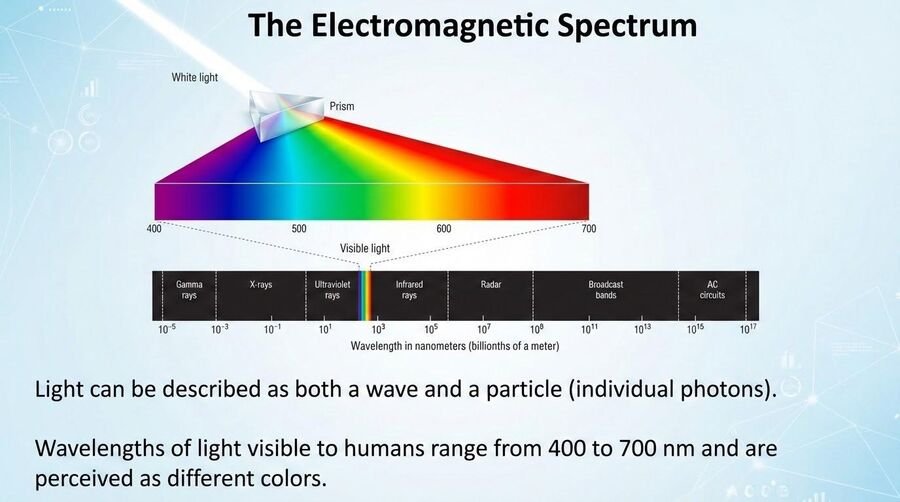
Two things are worth noticing about that sliver. First, light behaves like sound in one useful way — both are waves with a measurable wavelength and amplitude — but light is also a stream of particles (photons), which is why a single photon can, in principle, trip a receptor. Second, "visible" is a fact about us, not about light. Within our roughly 400–700 nanometer window, short wavelengths look violet and blue, long ones look red. Other animals draw the window somewhere else. Honeybees and many butterflies see into the ultraviolet, where flowers carry landing-strip patterns invisible to us; the kestrel hovering over a field tracks the UV-reflecting urine trails of voles. Pit vipers have a second, heat-sensing organ that detects infrared from warm prey in total darkness. There is nothing special about the band we happen to see. It's the band that a daylight-hunting primate's ancestors found worth the receptors.
The eye: getting light onto the retina
Light entering the eye first passes through the cornea, the clear outer layer that does most of the initial bending (refraction) of light rays. It then passes through the pupil, an opening whose size is controlled by the surrounding iris — this is the system that regulates how much light gets in, widening in dim conditions and constricting in bright ones. Behind the pupil, the lens fine-tunes the focus through a process called accommodation: muscles change the lens's shape to bend light precisely enough that it forms a sharp image on the back of the eye. All of that is just optics — getting a focused image onto the right surface. The actual sensory transduction, converting light energy into neural signals, happens at that back surface: the retina.
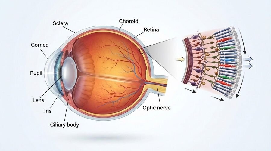
The retina isn't a uniform sheet — it contains two distinct types of photoreceptor, and the differences between them explain a lot of ordinary visual experience, from why you can't read street signs in your peripheral vision to why a dim room looks colorless.
Rods
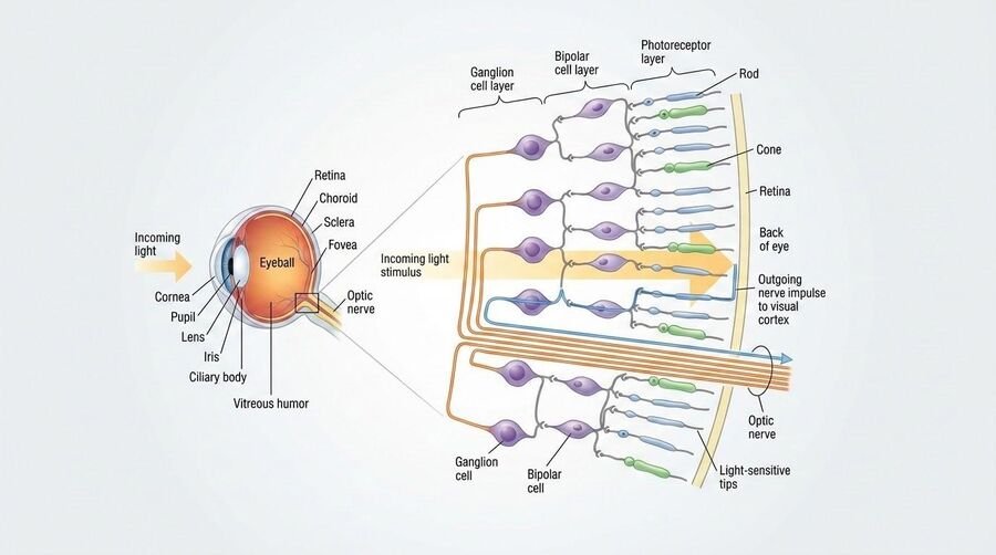
Highly sensitive to light, which makes them the workhorses of dim-light and nighttime vision. They're spread across the periphery of the retina rather than clustered in the center, which is why peripheral vision is better at detecting motion and low light than at resolving detail. Rods don't distinguish color — they contribute to vision in shades of gray.
Cones
Responsible for color vision and fine detail, but they need much more light to function well, which is why colors wash out in low light. Cones are concentrated in the fovea, the small central pit of the retina — exactly where you're pointing your gaze when you look directly at something to see it clearly.
The division of labor between rods and cones also explains why your eyes adjust so slowly when you step from bright sun into a dark room. This dark adaptation unfolds in two stages. The cones recover their sensitivity fairly quickly, over the first several minutes, but they never become very sensitive in dim light. The rods adapt far more slowly — regenerating their light-sensitive pigment takes time — but eventually become dramatically more sensitive, which is why complete dark adaptation can take twenty to thirty minutes, and why your deepest low-light vision is rod vision, and therefore colorless. The reverse process, light adaptation, is nearly instantaneous and briefly uncomfortable: it's the wash of blinding white you get walking out of a matinee into the afternoon sun.
The blind spot — and why you never notice it
Every retina has one small region with no photoreceptors at all: the spot where the optic nerve gathers all the retinal output and exits the eyeball, heading toward the brain. Because there are no rods or cones there, that patch of your visual field is, technically, not being sensed — it's a genuine gap in the incoming data. You never notice it in ordinary life for a simple reason: your brain fills it in, extrapolating from the surrounding visual context so smoothly that the gap disappears from awareness. It's a small, everyday example of a bigger theme in this unit — a lot of what feels like a direct, complete picture of the world is actually a construction, patched together from partial and imperfect input.
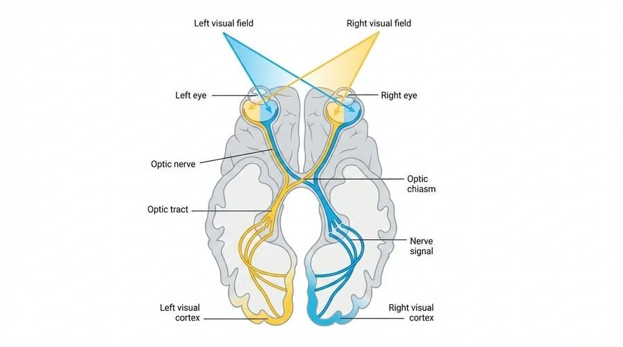
Since exam questions like to ask for the whole route, here it is in order. Light crosses the cornea, passes through the watery aqueous humor, enters through the pupil, is fine-focused by the lens, and travels through the gel-like vitreous humor that fills the eyeball to reach the retina — inverted and reversed, because a lens flips what it projects (the brain, having never known anything else, simply treats "up" as up). At the retina, light finally strikes the rods and cones. Transduction happens in their photopigments: in rods, a molecule called rhodopsin literally changes shape when a photon hits it, and that shape change kicks off the neural signal. (Rhodopsin is bleached by bright light and rebuilt slowly in the dark, which is the chemistry behind the slow half of dark adaptation you read about above; when rods can't do this efficiently, the result is night blindness.) Rods and cones pass their signals to bipolar cells, which pass them to ganglion cells, whose axons bundle together to form the optic nerve. In the fovea the wiring is nearly one cone per ganglion cell, which is why acuity is highest there; out in the periphery many rods converge on a single ganglion cell, trading detail for sensitivity. The two optic nerves meet at the optic chiasm, where the fibers carrying the right half of each eye's visual field cross to the left hemisphere and vice versa — so each side of the world is processed on the opposite side of the brain. From there the signal relays through the thalamus and arrives at the visual cortex in the occipital lobe at the back of the head, where feature detectors take over.
▲ A design flaw evolution never fixed
The vertebrate retina is wired "backwards." The optic nerve fibers and blood vessels that carry signals away from the photoreceptors sit in front of them, meaning light has to pass through a layer of neurons and vessels before it ever reaches the rods and cones doing the actual sensing. That's also exactly why the blind spot exists — it's the point where all those fibers have to bundle together and pass through the retina to exit the eye. This is a good illustration of a broader point about evolution: natural selection doesn't design from a blank slate for optimal engineering, it works with whatever arrangement history handed it, patching function onto structure rather than the other way around. Tellingly, the octopus eye — which evolved image-forming vision completely independently from vertebrates — is wired the "right" way round, with photoreceptors facing the light directly and no equivalent blind spot. Two separate evolutionary paths arrived at the same basic camera-eye design, but only one carried the wiring quirk, because only one was constrained by that particular ancestral starting point.
Feature detectors: perception built from parts
Once a signal leaves the retina and reaches the visual cortex, something remarkable happens at the level of individual neurons. Hubel and Wiesel (1962) recorded from single cells in the visual cortex and found that many neurons don't respond to light in general — they respond selectively to very specific features: an edge at one particular orientation, a line moving in one particular direction, and not others. These feature detectors effectively break the visual scene down into simple components — edges, angles, movement — before the brain reassembles those components into the complex, unified scene you actually experience. This work, which earned Hubel and Wiesel a Nobel Prize, is direct physiological evidence for something that might otherwise sound like pure theory: perception isn't a single act of "seeing," it's an assembly process, built up from many simple, specialized analyses happening in parallel.
Beyond individual feature detectors, the visual system divides its labor into two broad processing streams once information leaves the primary visual cortex. A ventral stream, running down toward the temporal lobe, handles what you're looking at — recognizing objects, faces, and words. A dorsal stream, running up toward the parietal lobe, handles where things are and how to act on them — guiding reaching, grasping, and movement through space (Goodale & Milner, 1992). The dissociation can be startling in patients with damage to just one stream: some can accurately reach out and grasp an object whose shape and orientation they cannot consciously describe, while others can describe an object in detail yet fumble badly when reaching for it. Seeing, it turns out, is not one unified act but at least two parallel jobs, handled by different neural real estate.
Color vision: two theories, both right
For a long time, color vision looked like a dispute with only one correct answer. It turns out both classic theories are correct — they just describe two different stages of the same system.
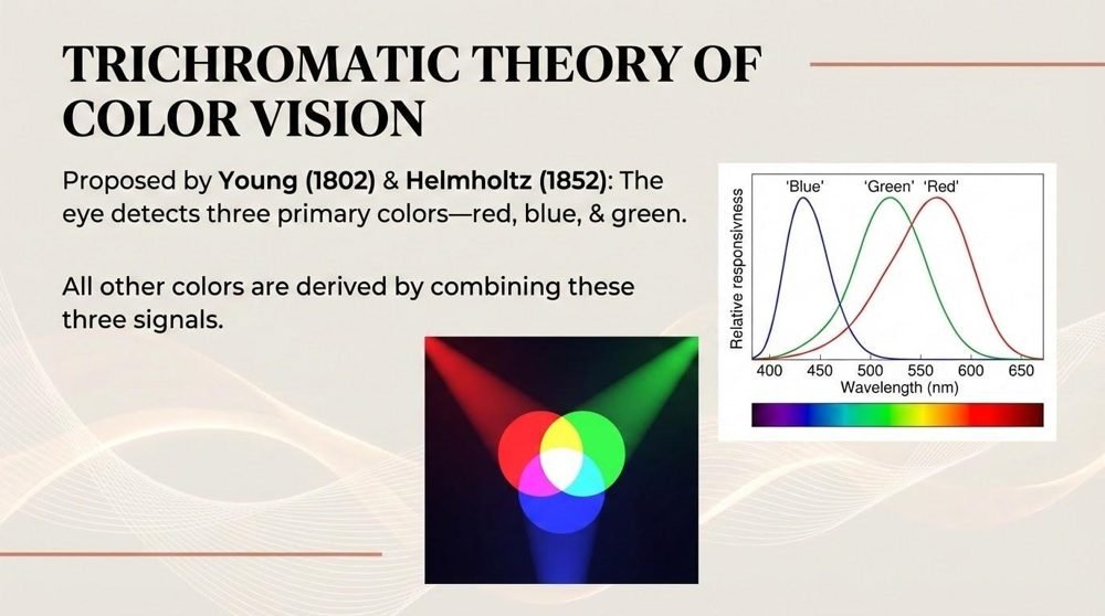
Trichromatic theory (Young–Helmholtz)
Proposes three types of cones, each maximally sensitive to a different range of wavelengths, and color perception arising from the combined pattern of activity across all three. This theory does an excellent job explaining what's happening at the receptor level — it correctly predicts things like which wavelength combinations will look identical to the eye, because it's grounded in the actual physical hardware of the retina.
Opponent-process theory (Hering)
Proposes that color is coded later, in opponent pairs — red versus green, blue versus yellow, and black versus white — where activating one member of a pair suppresses the other (Hering, 1920/1964). This theory explains things trichromatic theory can't reach on its own: why no color ever looks "reddish-green" or "bluish-yellow" to you, and why staring at a saturated color produces a negative afterimage in the opponent color once you look away.
◆ Try it: the afterimage
Stare at a strongly saturated red image for twenty or thirty seconds without looking away, then shift your gaze to a plain white or gray surface. You'll briefly see a green afterimage — not because anything green was ever presented, but because the red-green opponent channel in your visual system was pushed hard toward "red" and rebounds toward "green" once the stimulation stops. Trichromatic theory, by itself, has no mechanism for this. Opponent-process theory predicts it directly.
So the resolution to "which theory is right" isn't a compromise — it's a sequence. Trichromatic coding happens first, at the retina, where three cone types register wavelength. Opponent-process coding happens next, further along the visual pathway, recoding that trichromatic signal into red-green, blue-yellow, and black-white channels. Both theories are describing real, measured stages of the same system; they were never actually in competition once the full pathway was understood.
This two-stage system also explains the most common form of color blindness. Most color vision deficiencies are red-green deficiencies, tied to atypical or missing red- or green-sensitive cone photopigments, and the genes involved are carried on the X chromosome. Because of that X-linked inheritance pattern, red-green color blindness is substantially more common in males, who have only one X chromosome and so only need one altered copy to be affected, than in females, who have two X chromosomes and are typically shielded by having a second, typical copy.
The vocabulary here is straightforward once you count cone types. A person with all three is a trichromat. A dichromat has only two working cone types, and the red–green variety is by far the most common: roughly 8% of men of European descent, and well under 1% of women, for exactly the X-linked reason above. (You can usually pass as fine — the OpenStax author who tells his own story only found out when his seven-year-old refused to let him leave the house in green pants, an orange shirt, and a brown tie; Spielman et al., 2020.) A monochromat has one cone type or none, and the rod-only form — true grayscale vision — is vanishingly rare, on the order of one in 100,000, and comes packaged with poor acuity and painful sensitivity to bright light, since rods were never built to run the daytime shift. The standard screening tool is the Ishihara test: plates of colored dots hiding a number that pops out for trichromats and dissolves into the background for someone missing the relevant cone.
Color is made, not found
◆ Color doesn't live in the light
It's tempting to think of color as a property that objects and light simply have, the way an object has a certain weight. It's more accurate to say color is the brain's interpretation of wavelength information, and that interpretation is shaped heavily by surrounding context — the same wavelengths reaching your eye can be perceived as different colors depending on the lighting and colors around them. The internet-famous photo of "the dress" that different viewers confidently described as blue-and-black or white-and-gold became a cultural moment precisely because it exposed this normally invisible process: two people can receive the same physical input and have their visual systems construct genuinely different color experiences from it, depending on assumptions each brain was making about the lighting in the scene.
The myth
Color is a property of objects themselves — a ripe tomato simply "is" red, the same way it "is" round.
What's actually true
Objects don't emit or contain color — they selectively reflect certain wavelengths of light and absorb others. A tomato reflects long wavelengths and absorbs the rest; "red" is what your visual system constructs from that reflected wavelength, under a given set of lighting conditions. Change the light dramatically enough, or change the surrounding context, and the same physical tomato can be made to look like a different color, because the color was never a fixed property of the tomato to begin with.
Bodhi says
Notice the pattern connecting the blind spot and the dress: your visual system is constantly filling gaps and resolving ambiguity using assumptions you never consciously chose. Most of the time those assumptions are so reliable you never catch them working. Vision only reveals itself as a construction when something — a missing patch of retina, an ambiguous photo — pushes it into view.
Synesthesia: real cross-talk, not metaphor
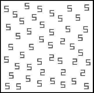
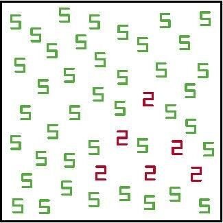
Most people experience the senses as cleanly separated — sound stays sound, color stays color. In synesthesia, that separation is incomplete: stimulation in one sensory or cognitive channel reliably and involuntarily triggers an experience in another. A common form is grapheme-color synesthesia, in which specific letters or numbers consistently evoke specific colors — the letter "A" might always appear tinged red to that person, every time, without effort or choice. This isn't imagination, exaggeration, or a poetic figure of speech. Ramachandran and Hubbard (2001) reviewed evidence that synesthesia is a genuine perceptual phenomenon, involuntary and remarkably consistent within a given person over time, and argued it likely reflects atypical cross-activation between neighboring brain regions that would ordinarily process these dimensions separately. Synesthesia is worth holding onto as a closing case for this unit: it's a vivid demonstration that what we call "sensation" is a set of separable neural channels, and that the usual boundaries between them are a feature of typical wiring, not a law of nature.
Check yourself
The one thing to carry out of this unit
Vision feels immediate and effortless, but almost nothing about it is simple or direct. Light gets bent and focused before it's even sensed; the retina that senses it is wired in a way evolution never bothered to optimize; individual cortical neurons are tuned to fragments of the scene rather than the whole picture; and color — maybe the most "obviously real" property of what you see — turns out to be assembled in two separate stages and then reshaped by context rather than read straight off the light itself. The throughline for this whole unit is the same one that will carry into perception: your visual experience is not a recording of the world, it's a construction the brain builds, usually so well that you never notice the work being done.
References
Goldstein, E. B., & Cacchione, T. (2021). Sensation and perception (11th ed.). Cengage.
Goodale, M. A., & Milner, A. D. (1992). Separate visual pathways for perception and action. Trends in Neurosciences, 15(1), 20–25. https://doi.org/10.1016/0166-2236(92)90344-8
Hering, E. (1964). Outlines of a theory of the light sense (L. M. Hurvich & D. Jameson, Trans.). Harvard University Press. (Original work published 1920)
Hubel, D. H., & Wiesel, T. N. (1962). Receptive fields, binocular interaction and functional architecture in the cat's visual cortex. The Journal of Physiology, 160(1), 106–154.
Ramachandran, V. S., & Hubbard, E. M. (2001). Synaesthesia—A window into perception, thought and language. Journal of Consciousness Studies, 8(12), 3–34.
Spielman, R. M., Jenkins, W. J., & Lovett, M. D. (2020). Psychology 2e. OpenStax. https://openstax.org/details/books/psychology-2e